
This is the first time in ten years that I haven't had an exam around summer-time. It feels odd, everyone around me is either exam-stressed or post-exam-relaxed, it's turning to summer and there's a definite final term feeling but this time I'm not really part of it. It's been an interesting year this year, since January I've not been involved in any part of research science, other than writing about it.
However luckily I'm still surrounded by lectures, seminars, talks and various other interesting stuff which no one seems to mind me occasionally turning up too. Seeing as I haven't written much about virus's lately I headed over to a talk the other day about Marek's disease, which is caused by a Herpes Virus and has a rather devastating affect on chickens.
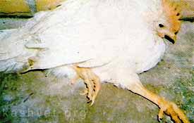
It makes them stand like this :(
It started back in the sixties, when rather a lot of chickens suddenly started dropping dead, sometimes up to 50% of all the stock in a large barn. Bear in mind these weren't the happy pecking-around-shrubs-of-grass chickens that feature on the front of free-range eggs, but rather a lot of chickens quite closely packed inside a big barn. The cause was found to be MDV - Marek's disease virus. After a lot of work a vaccination was found and given to all the chickens. Over $2 billion was saved by this, and the chickens were able to survive, right up until they got slaughtered for food.
But then, around the 1980s the disease suddenly reared it's head again, this time in a far more virulent form imaginatively labelled vvMDV (which stands for very virulent MDV). More research, another vaccine, and the deaths stopped.
Until just before 2000 when the virus evolved
again into an even
more virulent strain called vv+MDC, which means exactly what you think it does. Another vaccine was made (called Rispens) but at this point it was becoming fairly clear that this virus was behaving oddly. Three times it had changed, becoming more and more deadly each time:

Image from the presentation slides showing the development of new strains with an increase in virulence.
This is not normal behaviour. Virus's rely on their hosts, they can't replicate, survive or do
anything without a host cell, which means they have a vested interest in keeping the host alive. If anything, viral strains should evolve to become less virulent; a virus that kills the host will be a virus without a host and is therefore less likely to survive and propagate than a virus with a host.
It turns out in this case that the evolution of increase virulence is down exclusively to the way the vaccine interacts with the virus. The virus works by being inhaled into the chickens lungs, getting into the cells of the immune system (B and T cells) and causing a latent infection of the lymphocytes (T cells). Virus cells can also work their way to the epithelial cell of the feather follicles and will shed from under the feathers, thus keeping the virus in circulation.
In an ideal situation a vaccine would produce what is known as "sterilising immunity", where use of the vaccine kills all viruses dead. This is how almost all human vaccines work. With the Marek's disease vaccine however, the virus was not killed completely, but could still replicate and shed from the feathers. This means that the virus was still within the system, able to change and evolve. The vaccine however, does make the virus less likely to spread, which means that a more virulent form that the vaccine does
not protect against is able to outcompete the less virulent strain. Because the chickens are all in very close proximity, and because there are a lot of them, the more virulent strain can spread much faster throughout the population. With normal chickens, in small isolated populations this would not happen as a virus that virulent would run out of host and die out.
This creates a paradox - vaccines are needed to stop the chickens getting the virus, but at the same time use of the vaccine is creating an evolutionary environment in which a more virulent virus can grow. There are some responses to avoid this though. Firstly, to develop a virus that produces sterilising immunity, i.e that kills all thee virus dead. Secondly, to allow the chickens more room and freedom to stop the virulent strains spreading so quickly. And while this second solution sounds like every animal-rights campaigner's dream, remember that it only really works if very few people in the world eat chicken. In reality there are lots of people, and a limited space for chickens - eating chickens which have lead a happy healthy life is a privilege for a few, not the reality for the majority.
---
Witter RL (2001). Protective efficacy of Marek's disease vaccines. Current topics in microbiology and immunology, 255, 57-90 PMID: 11217428Witter RL (1997). Increased virulence of Marek's disease virus field isolates. Avian diseases, 41 (1), 149-63 PMID: 9087332---
Follow me on
Twitter!


